

Breast Center
At the OLYMPION General Clinic of Patra, an ultra modern Breast Center operates since November 2017, with holistic approach to breast conditions
This is accomplished with the cooperation of of different Specialties and Departments. More specifically, the Radiology laboratory contributes to Imaging (digital tomosynthesis, High- resolution breast ultrasound with elastography, MRI Scan) in the detection and diagnosis of breast conditions.

The Surgical Department consisting of experienced breast surgeons contributes to the clinical examination, the calculation of the risk, the appropriate and necessary in each case Surgical Procedures (i.e. biopsies, tumor excisions, mastectomies) and the surveillance.
The Pathology Laboratory contributes with the histological diagnosis and prognosis of breast conditions.
The Oncology Department contributes with the further treatment of patients, when needed (chemotherapy, hormonal therapy)..
The specialised physicians collaborating with the Breast Center, carry out a scientific council, every other week, in order to discuss the incidents and make decisions on the treatment approach for each patient.
The Breast Center is also supported by the Department of Medical Physics-radiophysics (for the radiation protection of the examinees and personnel, as well as the quality control of ionizing Imaging Systems and non-ionizing radiation), biomedical technology, and the Department of Medical Informatics.
The Center’s aim is to offer in a single visit, as long as the examinee agrees, diagnostic exams (clinical exam, mammography, ultrasound), and if required, biopsy and histological exam.
More specifically, the following medical services are provided at the OLYMPION Breast Center:
Preventive examination (Screening) – Diagnosis
- Digital tomosynthesis of Breast (3d mammogram)
- High Resolution breast ultrasound
- Breast elastography (type shearwave, SWE)
- Vascular breast Ultarsound
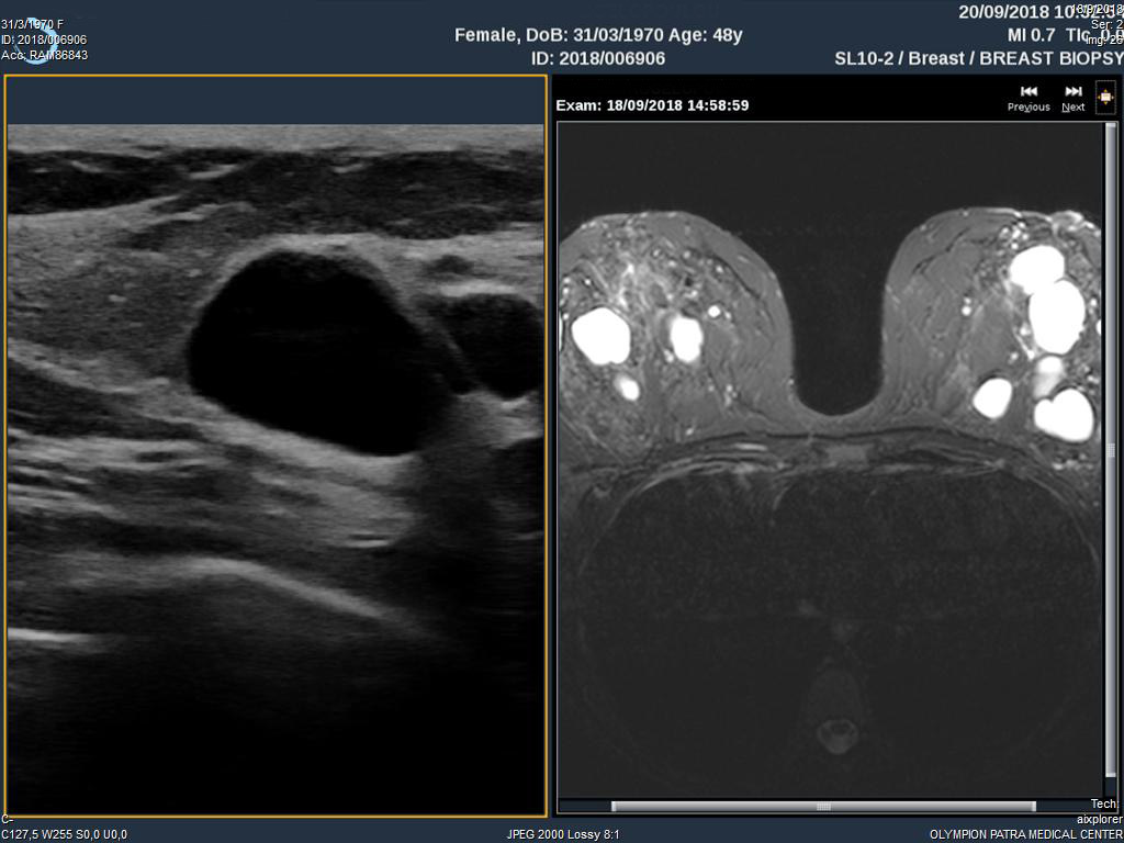
- Breast MRI
Exams are archived electronically
Diagnostic test – Holistic approach
- Breast MRI
- Lesions detection with wire guide
- Imaging guided Biopsy
- Marking lesions with clips before starting chemotherapy
- Clinical exam by a specialized Mastologist
- Oncology Assessment
- Surgery
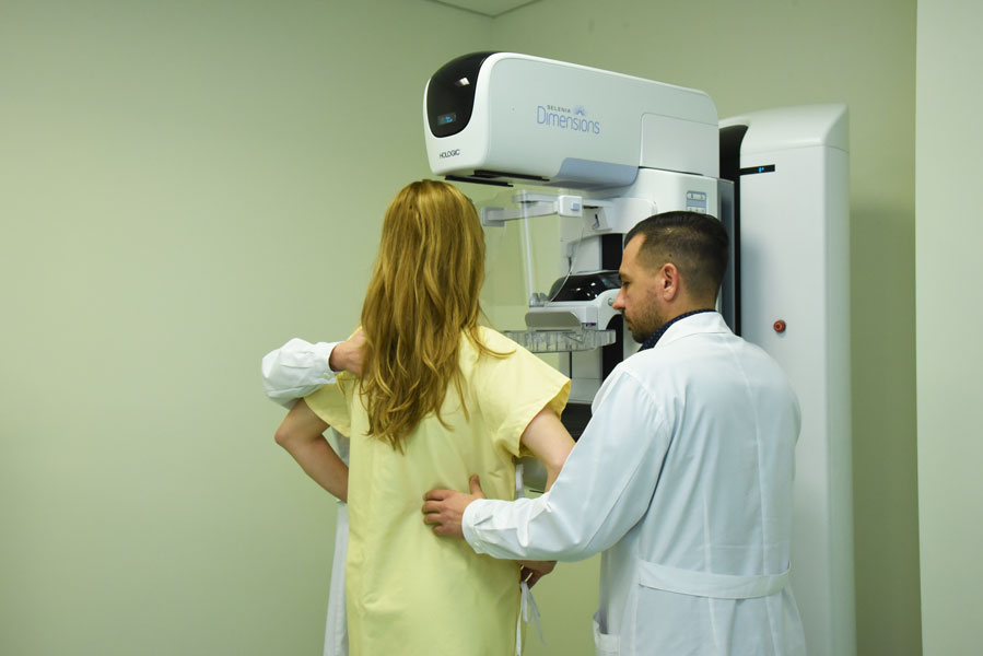
Digital tomosynthesis System (3D Mammogram, Selenia Dimensions, Hologic)
One of the most important "medical tools" at the Breast Center is the digital breast tomosynthesis system (selenia dimensions, 3d mammography), constructed by Hologic. It is the first Digital Breast Tomosynthesis system installed in the private sector in western Greece.
Breast Tomosynthesis – the Future in Preventive examination (Screening)
The Breast Tomotomthesis system is the latest technological development in the depiction of breast conditions, since the automatic rotation of the lamp allows the 3d imaging of the breasts, i.e. an upgrated exam compared to the Digital (2d) mammography.
Tomosynthesis (3D)-Operating principle
During the rotation of the bulb, 15 low-dose views (images) are taken, that with the appropriate software are resynthesized in slices that are 1 millimeter apart, giving the radiologist the ability to study the breast in detail, revealing areas that were concealed due to projection in the 2D images.
The technique, in terms of the method that the exam is carried out and the placement of the examinee, is not different from the technique of 2D Mammography, simply the lamp takes pictures at different angles (-7.50 to + 7.50) in a short time (3.7 sec). This system has the potential for 2D Digital Mammogram, for 3D Digital Mammography (tomosynthesis), as well as a combination of these (2d + 3d Digital Mammogram). Finally, this system has the potential of producing synthesized 2D digital mammography, which are created by the digital breast tomosynthesis, without additional charge in dosage, for the examinee (2D synthesized + 3D Digital Mammography).
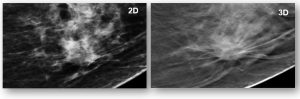
Why 3D digital mammogram?
The digital tomosynthesis allows the distinction of lesions that due to tissue projection are indistinguishable or not distinct in the 2D Digital mammogram, i.e. it achieves clearer detection (as it provides the possibility of distinguishing the lesions boundaries) and diagnosis (as it provides the possibility of distinguishing benign from malignant lesions). According to recent published studies, the benefits from the 3D Digital Mammogram are the following:
- 40% increase in the detection of invasive cancers
- 27% increase in the detection of cancers (invasive and non-invasive)
- 15% reduction in false positive results.
Low dose mammogram with use of synthetic images (Synthesized 2D Digital Mammography)
The OLYMPION Breast Center is one of the 3 laboratories in Greece with Digital Mammography constructed by HOLOGIC, which has been installed and operates and disposes production software of synthetic 2D Digital Mammography (Synthesized 2D mammography, C-VIEWTM) , which is the evolution of the breast tomosynthesis technique.
The advantages of using Synthetic 2D Digital Mammography are the following :
- Through the revolutionary C-VIEWTM technique, the need for 2D Digital Mammography is completely eliminated, as the 3D imaging (tomosynthesis) creates the synthetic 2D image of the same breast.
- The synthetic 2d image (synthesized 2D, "C-VIEW") results from appropriate processing of contrast enhancement and presents with sharper definition, all the useful information and Structures of a 2D examination.
- The total dose and time, of a complete mammographic exam are significantly reduced.
- The synthetic 2D image (synthesized 2D, "C-VIEW") achieves a better display of linear structures and radial projections that are often found in masses and architectural disorders, in relation to the 2D image.
- The combination of tomosynthesis images and synthetic image, detects more masses and architectural disorders, compared to the 2D digital mammography.
3D mammography exam with HOLOGIC (Selenia Dimensions)
With regard to the 3D mammographic exam with the HOLOGIC (Selenia Dimensions) construction House system :
- It is certified as a superior exam to the simple 2D Digital mammography, according to the food and drug administration organization (FDA).
- It has been proven to be superior to the 2D Digital mammography for women with dense breast (FDA approval).
- The combination of 3D breast tomosynthesis and 2D Digital Mammography (3D+2D) is clinically proven to be more accurate (increase of sensitivity and reduction of the recall numbers) than the individual use of 2D Digital Mammography, for all types of breast.
- The combination of 3D Digital breast tomynthesis and synthesized 2D Digital Mammography (3D + synthesized 2D) has a higher performance in traceability than the individual use of 2D Digital Mammography.
- The combination of 3D Digital breast tomosynthesis and synthesized 2D Digital mammography (3D + synthesized 2D) contributes to a lower dose burden of the examinee compared to the combination of the 3D breast tomography and 2D Digital mammography (3D+ 2D).
High Definition Ultrasound
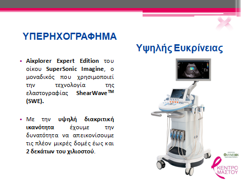
The breast center has been equipped with the ultrasound system Aixplorer Expert Edition of the Super Sonic Imagine construction House, high resolution capacity where small structures are depicted (up to 0.25 mm), and the only one using the technology of Elastography shearwave (shear wave TM Elastography, SWE). This technology quantifies reliably and repeatedly the mechanical and elastic properties of the tissues and is complementary to the conventional ultrasonography (B-Mode and color Doppler) for making the final decision in the diagnosis.
It has been proved to be an additional supply for the doctor-Radiologist, in the improvement of management of patients with breast lesions. In particular, it achieves (a) increase in the reliability of risk assessment of cancer based on ultrasound (ultrasound cancer risk Assessment) and (b) increase in the diagnostic accuracy of breast cancer with increased sensitivity and reduction of false positive results. It has also been shown to contribute pre-operatively in the assessment of the size of breast cancer, cancer aggression and prognosis. Finally, it is possible to contribute to the control and prediction of the response of chemotherapy in cases where the cancer has not been removed.
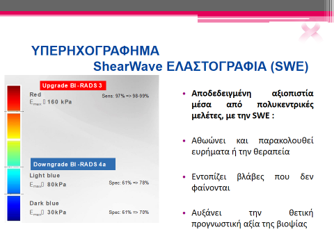
ULTRASOUND SHEARWAVE ELASTOGRAPHY (SWE)
Proven credibility through polycentric studies, with the SWE:
- Acquits and observes findings or the treatment
- Localizes lesions that are not apparent
- Increases the positive prognostic value of the biopsy
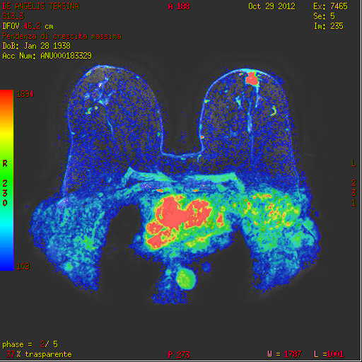
Magnetic Mammography
What is it?
~ It's a magnetic resonance imaging (MRI), focused on breast study.
~ It is complementary to mammography and ultrasound and does not substitute them.
~ It is the most sensitive method today for breast study (90%).
~ It can detect lesions at an early stage, compared to mammography and ultrasound.
~ It has a large negative predictive value (> 98%).
~ Ideally performed on the 7th -12th day of the cycle.
~ It's a painless method.
~ Non-ionized radiation.
Indications:
- It is an exam for women of high risk for breast cancer development.
- For the assessment of the other breast of women with known breast cancer.
- For women with implants or previous surgery.
- To illustrate the extent of the disease in newly diagnosed cases.
- To assess the response to chemotherapy.
- For further investigation of suspicious findings in the mammography, the clinical exam or the ultrasound, and for specific types of cancer (lobular carcinoma, unknown primary carcinoma ).
Frequently Asked Questions - Answers
- What day of the cycle do I test?
The preventive annual check is ideally done in the 2nd week of the cycle (7th -14th day). The breast MRI is performed on the 7th -12th day. If you are receiving hormonal therapy, you should tell your doctor. If you have a symptom, contact your doctor, do not delay the exam. - Do I need to submit previous exams?
It is necessary, as studies have shown that by submitting the previous exams, a large proportion of the findings initially placed under close observation, are finally acquitted. - I have a small breast. Should I have a mammogram?
All women over 40 years old need to have a mammogram annually. - I have implants. Do I have a mammogram?
There are special imaging techniques. Women > 40 with implants need to have a mammogram annually.
- Do I always have to do ultrasound with the yearly mammogram?
It will be necessary if you have dense breasts or if the radiologist needs to define a finding. - I'm just going to have an ultrasound to avoid radiation?
breast cancer in the general population. The reduction of mortality due to early diagnosis with mammography, is 40%. Mammography is the only diagnostic method that has been shown to reduce the risk and mortality. Ultrasound is not a screening method on its own. Ultrasound is indicated in young women as a method of preventive exam. Many times it will be necessary with your mammogram as a complementary exam. - What is a second-glance ultrasound?
It is an ultrasound focused on the findings of the breast MRI or the mammography. At the OLYMPION Breast Center, parallel imaging-linking of images is possible.